
» Zoom » Download
![]()
Original filename: Stenaulorhynchus NHMUK PVR 36618 T2 5x RTL w scale LAGs directions MAma MB.jpg
Morphobank media number
M346740
Taxonomic name
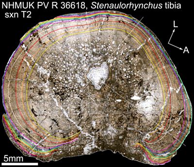
† Stenaulorhynchus stockleyi Haughton, 1932
Specimen
† Stenaulorhynchus stockleyi Haughton, 1932 (NHMUK/PV:R36618)
Specimen notes
Partial skeleton including rostrum (mostly R maxilla and R dentary, but also including mandibular symphysis and anteriormost portion of L dentary), 14 vertebrae, rib, partial left and right humeri, left femur, right and left tibiae, right and left calcanea.
View
tibia - mid-diaphyseal cross section
Media loaded by
Sarah Werning
Copyright holder
The Natural History Museum, London (Image taken and edited by Sarah Werning)
Copyright information
Permission to use media on MorphoBank granted by copyright holder
Media notes
Tibia, mid-diaphysis, transmitted light with a single plane polarizer. Colored lines correspond to growth marks; these follow the same color pattern as in the femur. Solid white lines spanning the diameter represent major and minor axes for the cross section. The white line spanning only the radius in the element (posterolateral quadrant) is the transect along which the outermost zones were measured.
Scale = 5mm. Abbreviations: A, anterior; L, lateral.
Section T2.
Vertical "striping" results from image overlap during the photomontaging process.
Media loaded on
November 30 2014 at 19:10:04
This media record has been viewed 741 times ![]()