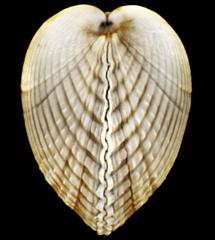
Project 3107: A. Ziegler, C. Bock, D. R. Ketten, R. W. Mair, S. Mueller, N. Nagelmann, E. D. Pracht, L. Schröder. 2018. Digital three-dimensional imaging techniques provide new analytical pathways for malacological research. American Malacological Bulletin. 36 (2):248-274.
Abstract
Research on molluscan specimens is increasingly being carried out using high-throughput molecular techniques. Due to their efficiency, these technologies have effectively resulted in a strong bias towards genotypic analyses. Therefore, the future large-scale correlation of such data with the phenotype will require a significant increase in the output of morphological studies. Three-dimensional (3D) scanning techniques such as magnetic resonance imaging (MRI) or computed tomography (CT) can achieve this goal as they permit rapidly obtaining digital data non-destructively or even entirely non-invasively from living, fixed, and fossil samples. With a large number of species and a relatively complex morphology, the Mollusca would profit from a more widespread application of digital 3D imaging techniques. In order to provide an overview of the capacity of various MRI and CT techniques to visualize internal and external structures of molluscs, more than twenty specimens ranging in size from a few millimeters to well over one meter were scanned in vivo as well as ex vivo. The results show that all major molluscan organ systems can be successfully visualized using both MRI and CT. The choice of a suitable imaging technique depends primarily on the specimen's life condition, its size, the required resolution, and possible invasiveness of the approach. Apart from visual examples derived from more than two dozen scans, the present article provides guidelines and best practices for digital 3D imaging of a broad range of molluscan taxa. Furthermore, a comprehensive overview of studies that previously have employed MRI or CT techniques in malacological research is given.Read the article »
Article DOI: 10.4003/006.036.0205
Project DOI: 10.7934/P3107, http://dx.doi.org/10.7934/P3107
| This project contains |
|---|
Download Project SDD File |
Currently Viewing:
MorphoBank Project 3107
MorphoBank Project 3107
- Creation Date:
13 February 2018 - Publication Date:
24 January 2019 - Media downloads: 201

Authors' Institutions ![]()
- Harvard University
- Universitaet Muenster (Westfälische Wilhelms-Universität Münster)
- Alfred-Wegener-Institut
- Charité – Universitätsmedizin Berlin
- Deutsches Zentrum für Neurodegenerative Erkrankungen (DZNE)
- Leibniz-Forschungsinstitut für Molekulare Pharmakologie (FMP)
- Rheinische Friedrich-Wilhelms-Universität Bonn (University of Bonn)
- Woods Hole Oceanographic Institution
Members
| member name | taxa |
specimens |
media | media notes |
| Alexander Ziegler Project Administrator | 19 | 20 | 103 | 23 |
| Christian Bock Full membership | 0 | 0 | 0 | 0 |
| Darlene R. Ketten Full membership | 0 | 0 | 0 | 0 |
| Ross Mair Full membership | 0 | 0 | 0 | 0 |
| Susanne Mueller Full membership | 0 | 0 | 0 | 0 |
| Nina Nagelmann Full membership | 0 | 0 | 0 | 0 |
| Eberhard D. Pracht Full membership | 0 | 0 | 0 | 0 |
| Christina L. Sagorny Full membership | 0 | 0 | 0 | 0 |
| Leif Schröder Full membership | 0 | 0 | 0 | 0 |
| Gillian Trombke Full membership | 0 | 0 | 0 | 0 |
Project has no matrices defined.
Project downloads 
| type | number of downloads | Individual items downloaded (where applicable) |
| Total downloads from project | 447 | |
| Media downloads | 201 | M480210 (4 downloads); M480212 (3 downloads); M480182 (2 downloads); M475712 (7 downloads); M480243 (2 downloads); M475711 (3 downloads); M475713 (3 downloads); M475714 (3 downloads); M475715 (2 downloads); M475716 (4 downloads); M475717 (2 downloads); M475718 (4 downloads); M475719 (3 downloads); M475720 (4 downloads); M475721 (2 downloads); M475722 (2 downloads); M475723 (4 downloads); M475724 (6 downloads); M475725 (2 downloads); M475726 (2 downloads); M475727 (2 downloads); M475728 (2 downloads); M475729 (4 downloads); M475730 (3 downloads); M475731 (4 downloads); M475732 (7 downloads); M475733 (4 downloads); M475734 (3 downloads); M480166 (3 downloads); M480167 (3 downloads); M480168 (2 downloads); M480169 (2 downloads); M480170 (1 download); M480171 (2 downloads); M480172 (1 download); M480173 (1 download); M480174 (2 downloads); M480175 (2 downloads); M480176 (1 download); M480177 (1 download); M480178 (1 download); M480179 (1 download); M480180 (1 download); M480181 (1 download); M480183 (1 download); M480184 (1 download); M480185 (1 download); M480186 (3 downloads); M480187 (1 download); M480188 (1 download); M480189 (1 download); M480190 (1 download); M480191 (1 download); M480192 (1 download); M480193 (1 download); M480194 (1 download); M480195 (2 downloads); M480196 (1 download); M480197 (1 download); M480198 (1 download); M480199 (1 download); M480200 (3 downloads); M480201 (3 downloads); M480202 (3 downloads); M480203 (2 downloads); M480204 (5 downloads); M480205 (3 downloads); M480206 (1 download); M480207 (1 download); M480208 (2 downloads); M480209 (2 downloads); M480211 (2 downloads); M480213 (1 download); M480214 (1 download); M480215 (2 downloads); M480216 (1 download); M480217 (1 download); M480218 (1 download); M480219 (2 downloads); M480220 (1 download); M480221 (1 download); M480222 (1 download); M480224 (1 download); M480225 (2 downloads); M480226 (1 download); M480227 (1 download); M480228 (1 download); M480229 (1 download); M480230 (1 download); M480231 (1 download); M480232 (1 download); M480233 (1 download); M480234 (1 download); M480235 (1 download); M480236 (1 download); M480237 (2 downloads); M480238 (2 downloads); M480239 (1 download); M480240 (1 download); M480241 (1 download); M480242 (1 download); M480244 (1 download); M480245 (1 download); |
| Project downloads | 230 | |
| Document downloads | 16 | DTI data Sepia officinalis (16 downloads); |

