
Project 3517: M. T. Silcox, G. F. Gunnell, J. I. Bloch. 2020. Cranial anatomy of Microsyops annectens (Microsyopidae, Euarchonta, Mammalia) from the middle Eocene of Northwestern Wyoming. Journal of Paleontology. 94 (5):979-1006.
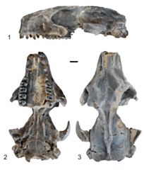
Specimen: † Microsyops annectens (Marsh, 1872) (UW:12362)
View: lateral, ventral and dorsal
View: lateral, ventral and dorsal
Abstract
The Microsyopidae are extinct mammals from the late Paleocene–late Eocene of North America and the late Paleocene of Europe. While results from phylogenetic analyses support euarchontan affinities, specific relationships ofmicrosyopids to other plesiadapiforms (plausible stem primates), Euprimates (crown primates), Scandentia (treeshrews), and Dermoptera (colugos) are unresolved. An exceptionally well-preserved cranium of Microsyops annectens includes a basicranium that is generally primitive relative to that of other extinct and extant euarchontans in having: (1) a transpromontorial groove for an unreduced internal carotid artery (ICA) entering the middle ear posteromedially; (2) grooves (not tubes) on the promontorium, marking the course for both stapedial and promontorial branches of the ICA; (3) a foramen faciale that opens into the middle ear cavity, with the facial nerve exiting through a stylomastoid foramen primitivum; and (4) unexpanded caudal and rostral tympanic processes of the petrosal. The absence of any preserved bullar elements in the middle ear contrasts with that of other plesiadapiforms for which the region has been recovered, all of which have evidence of an ossified bulla. Microsyops lacks many of the specialized cranial characteristics of crown scandentians and dermopterans. The basicranial anatomy of microsyopids does not provide evidence in support of a clear link to any of the extant euarchontans, and suggests that the primitive morphology of this region in Euarchonta was little differentiated from that observed in the primitive placental mammals.
Read the article »
Article DOI: 10.1017/jpa.2020.24
Project DOI: 10.7934/P3517, http://dx.doi.org/10.7934/P3517
| This project contains |
|---|
Download Project SDD File |
Currently Viewing:
MorphoBank Project 3517
MorphoBank Project 3517
- Creation Date:
19 August 2019 - Publication Date:
20 May 2020
This research
supported by
Authors' Institutions ![]()
- Duke University
- University of Florida
- University of Toronto
Members
| member name | taxa |
specimens |
media |
| Mary Silcox Project Administrator | 1 | 1 | 2 |
Project has no matrices defined.
Project downloads 
| type | number of downloads | Individual items downloaded (where applicable) |
| Total downloads from project | 136 | |
| Project downloads | 133 | |
| Document downloads | 3 | Matrix used to produce Figure 2 (modified from Bloch et al., 2016) (3 downloads); |