
Project 3700: T. L. Green, P. M. Gignac. 2020. Osteological description of casque ontogeny in the southern cassowary (Casuarius casuarius) using micro-CT imaging. The Anatomical Record. 2020 ( ):1–19.
Abstract
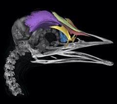
Extant cassowaries (Casuarius) are unique flightless birds found in the tropics of Indo-Australia. They have garnered substantial attention from anatomists with focus centered on the bony makeup and function of their conspicuous cranial casques, located dorsally above the orbits and neurocranium. The osteological patterning of the casque has been formally described previously; however, there are differing interpretations between authors. These variable descriptions suggest that an anatomical understanding of casque anatomy and its constituent elements may be enhanced by developmental studies aimed at further elucidating this bizarre structure. In the present study, we clarify casque osteology of the southern cassowary (C. casuarius) by detailing casque anatomy across an extensive growth series for the first time. We used micro-computed tomography (µCT) imaging to visualize embryonic development and post-hatching ontogeny through adulthood. We also sampled closely related emus (Dromaius novaehollandiae) and ostriches (Struthio camelus) to provide valuable comparative context. We found that southern cassowary casques are comprised of three paired (i.e., nasals, lacrimals, frontals) and two unpaired elements (i.e., mesethmoid, median casque element). Although lacrimals have rarely been considered as casque elements, the contribution to the casque structure was evident in µCT images. The median casque element has often been cited as a portion of the mesethmoid. However, through comparisons between immature C. casuarius and D. novaehollandiae we document the median casque element as a distinct unit from the mesethmoid.Read the article »
Article DOI: 10.1002/ar.24477
Project DOI: 10.7934/P3700, http://dx.doi.org/10.7934/P3700
| This project contains |
|---|
Download Project SDD File |
Currently Viewing:
MorphoBank Project 3700
MorphoBank Project 3700
- Creation Date:
17 April 2020 - Publication Date:
17 July 2020 - Media downloads: 3

This research
supported by
Authors' Institutions ![]()
- Oklahoma State University
- American Museum of Natural History (AMNH)
- MicroCT Imaging Consortium for Research and Outreach, AK
Members
| member name | taxa |
specimens |
media |
| Paul Gignac Project Administrator | 1 | 3 | 1 |
Project has no matrices defined.
Project downloads
| type | number of downloads | Individual items downloaded (where applicable) |
| Total downloads from project | 186 | |
| Project downloads | 160 | |
| Document downloads | 23 | Green & Gignac, 2020 - Supplementary Video File 1 -AMNH SKEL 963 FRONTAL (4 downloads); Green & Gignac, 2020 - Supplementary Spreadsheet 1 - ANATOMICAL RECORD (8 downloads); Green & Gignac, 2020 - Supplementary Video File 2 -AMNH SKEL 963 SAGITTAL (3 downloads); Green & Gignac, 2020 - Supplementary Video File 3 -AMNH SKEL 963 TRANSVERSE (2 downloads); Green & Gignac, 2020 - Supplementary Video File 4 -AMNH SKEL 962 FRONTAL (1 download); Green & Gignac, 2020 - Supplementary Video File 5 -AMNH SKEL 962 SAGITTAL (1 download); Green & Gignac, 2020 - Supplementary Video File 6 -AMNH SKEL 962 TRANSVERSE (4 downloads); |
| Media downloads | 3 | M692751 (3 downloads); |