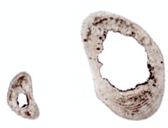
Project 4522: D. Nakai, A. Boyde. 2023. A new thin sectioning method for observation of higher resolution images in bone histomorphology. Journal of Vertebrate Paleontology. 43 (3):e2298402.
Abstract
Observing bone microstructure may reveal aspects of the ecological behavior and physiological mechanisms of extant and extinct vertebrates. High quality bone thin sections are required for superior resolution of histomorphological observation. We encountered three problems in studying bone thin sections of the material available to us: (1) opacity due to post-mortem migration of blood pigment; (2) birefringence of the epoxy resin used to embed the samples which disturbs observations in a polarizing microscope; and (3) abrasive powder residue which becomes incorporated into the bone sections. Modified procedures were developed to solve these problems. The blood pigment can be removed by N,N,N’,N’-tetrakis (2-hydroxypropyl) ethylenediamine. The epoxy resin can be removed by repetitive treatments with acetone and dichloromethane. Water-proof silicon carbide papers can be used for grinding and polishing thin sections instead of abrasive powder. These improvements make clearer, more transparent sections with improved images, which are easier to measure.Read the article »
Article DOI: 10.1080/02724634.2023.2298402
Project DOI: 10.7934/P4522, http://dx.doi.org/10.7934/P4522
| This project contains |
|---|
Download Project SDD File |
Currently Viewing:
MorphoBank Project 4522
MorphoBank Project 4522
- Creation Date:
10 January 2023 - Publication Date:
11 July 2024
Authors' Institutions ![]()
- Nagoya University
- Queen Mary University of London
Members
| member name | taxa |
specimens |
media | media notes |
| Daichi Nakai Project Administrator | 5 | 5 | 30 | 29 |

