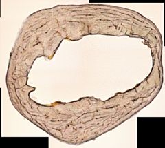
Project 5264: D. Nakai, Y. Yokohata. 2024. Bone microstructure as an indicator of digging ability in moles (Talpidae, Eulipotyphla). Journal of Anatomy. 245 (4):572–582.
Abstract
Talpid moles (Talpidae, Eulipotyphla) are mammals highly specialised in burrowing using their forelimbs. Fossoriality has allowed moles to expand their ecological niche by enabling access to subterranean resources and spaces. This specialisation in burrowing has led to adaptations in the forelimb bones of moles for humeral rotation digging, a distinctive strategy unparalleled among other diggers. While bone robustness has been examined in moles through external morphology, the adaptation of bone microstructure to digging strategy remains unclear. Based on two assumptions, (1) the humerus of moles is subjected to a torsional load due to humeral rotation digging, and (2) the magnitude of torsional load correlates with the compactness of the substrate in which the individuals can dig, we hypothesised that humeral rotation digging influences bone microstructure. Comparative analyses of transverse sections from the humeri and femora of three mole species (Mogera imaizumii, Mogera wogura and Urotrichus talpoides; Talpidae) and an outgroup eulipotyphlan (Suncus murinus; Soricidae) revealed that (1) vascular canals distributed in the humeri of moles align more predominantly circumferential along the bone walls, indicating an adaptation to the torsion generated by humeral rotation digging, and (2) the laminarity of vascular canals, particularly in Mogera species compared with Urotrichus, potentially reflects differences in the magnitude of load due to substrate compactness during digging. The aligned vascular canals are distinctive traits not observed in mammals employing other digging strategies. This suggests that vascular canal laminarity can be an indicator of not only humeral rotation digging in fossorial animals, but also the variation of eco-spaces in talpid species.Read the article »
Article DOI: 10.1111/joa.14114
Project DOI: 10.7934/P5264, http://dx.doi.org/10.7934/P5264
| This project contains |
|---|
Download Project SDD File |
Currently Viewing:
MorphoBank Project 5264
MorphoBank Project 5264
- Creation Date:
28 May 2024 - Publication Date:
25 November 2024
Authors' Institutions ![]()
- Nagoya University
- University of Toyama
Members
| member name | taxa |
specimens |
media |
| Daichi Nakai Project Administrator | 4 | 27 | 153 |

