Project 2083: C. Kolb, T. M. Scheyer, A. M. Lister, C. Azorit, J. de Vos, M. A. J. Schlingemann, G. Rössner, N. T. Monaghan, M. R. Sánchez-Villagra. 2015. Growth in fossil and recent deer and implications for body size and life history evolution. BMC Evolutionary Biology. 15 (19):1-15.
Abstract
BackgroundBody size variation within clades of mammals is widespread, but the developmental and life-history mechanisms by which this variation is achieved are poorly understood, especially in extinct forms. An illustrative case study is that of the dwarfed morphotypes of Candiacervus from the Pleistocene of Crete versus the giant deer Megaloceros giganteus, both in a clade together with Dama dama among extant species. Histological analyses of long bones and teeth in a phylogenetic context have been shown to provide reliable estimates of growth and life history patterns in extant and extinct mammals.Results
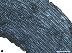
Similarity of bone tissue types across the eight species examined indicates a comparable mode of growth in deer, with long bones mainly possessing primary plexiform fibrolamellar bone. Low absolute growth rates characterize dwarf Candiacervus sp. II and C. ropalophorus compared to Megaloceros giganteus displaying high rates, whereas Dama dama is characterized by intermediate to low growth rates. The lowest recorded rates are those of the Miocene small stem cervid Procervulus praelucidus. Skeletal maturity estimates indicate late attainment in sampled Candiacervus and Procervulus praelucidus. Tooth cementum analysis of first molars of two senile Megaloceros giganteus specimens revealed ages of 16 and 19 years whereas two old dwarf Candiacervus specimens gave ages of 12 and 18 years.Conclusions
There is a rich histological record of growth across deer species recorded in long bones and teeth, which can be used to understand ontogenetic patterns within species and phylogenetic ones across species. Mean maximum growth rates plotted against the anteroposterior bone diameter as a proxy for body mass indicate three groups: one with high growth rates including Megaloceros, Cervus, Alces, and Dama; an intermediate group with Capreolus and Muntiacus; and a group showing low growth rates, including dwarf Candiacervus and Procervulus. Dwarf Candiacervus, in an allometric context, show an extended lifespan compared to other deer of similar body size such as Mazama which has a maximum longevity of 12 years in the wild. Comparison with other clades of mammals reveals that changes in size and life history in evolution have occurred in parallel, with various modes of skeletal tissue modification.
Read the article »
Article DOI: 10.1186/s12862-015-0295-3
Project DOI: 10.7934/P2083, http://dx.doi.org/10.7934/P2083
| This project contains |
|---|
Download Project SDD File |
Currently Viewing:
MorphoBank Project 2083
MorphoBank Project 2083
- Creation Date:
13 December 2014 - Publication Date:
16 February 2015 - Media downloads: 5

Authors' Institutions ![]()
- Museu de Ciències Naturals, Barcelona
- Canadian Food Inspection Agency
- Institut de Biologia Evolutiva (CSIC-UPF), Barcelona
Members
| member name | taxa |
specimens |
media | media notes |
| Christian Kolb Project Administrator | 0 | 1 | 1 | 0 |
| Maureen Admin Full membership | 0 | 0 | 0 | 0 |
| MorphoBank Curator Full membership | 9 | 15 | 17 | 0 |
Project has no matrices defined.
Project downloads 
| type | number of downloads | Individual items downloaded (where applicable) |
| Total downloads from project | 484 | |
| Document downloads | 39 | Figure S1. Femoral bone cortex of Dama and Procervulus. (14 downloads); Figure 2. Histological growth series of dwarf deer femora with skeletal reconstructions (modified from van der Geer et al. 2006) and specimens sampled (anterior view). (5 downloads); Figure 3. Cervid bone tissue and growth marks. (2 downloads); Figure 4. Bone cortex of Megaloceros giganteus. (14 downloads); Figure 5. Cervid bone remodelling. (2 downloads); Figure 8. Cervid tooth histology. (2 downloads); |
| Project downloads | 440 | |
| Media downloads | 5 | M374383 (4 downloads); M374238 (1 download); |

